Tendon Diagram Of Hand : Body Anatomy Upper Extremity Tendons The Hand Society - The tendons that run down our fingers are held in place by a series of.
Tendon Diagram Of Hand : Body Anatomy Upper Extremity Tendons The Hand Society - The tendons that run down our fingers are held in place by a series of.. The tendon travels along the inside of the forearm on the side of the small finger and crosses the wrist. Few structures of the human anatomy are as unique as the hand. Tendons allow fingers to pinch, grasp, grip, and straighten. In this image, you will find ulnar artery, ulnar nerve, common palmar digital branches of median nerve, hypothenar muscles, common flexor sheath, ulnar bursa, 5th finger tendinous sheath, synovial tendinous sheath, midpalmar space, insertion of flexor digitorum superficialis tendon, insertion of flexor. They are controlled by muscles in the forearm.
Sechrest, md narrates an animated tutorial on. The carpals join with the two forearm bones, the radius and ulna, forming the wrist joint. What parts make up the hand 2. These joints are called the metacarpophalangeal joints(mcp joints). It also attaches to the trapezium, one of your wrist bones.
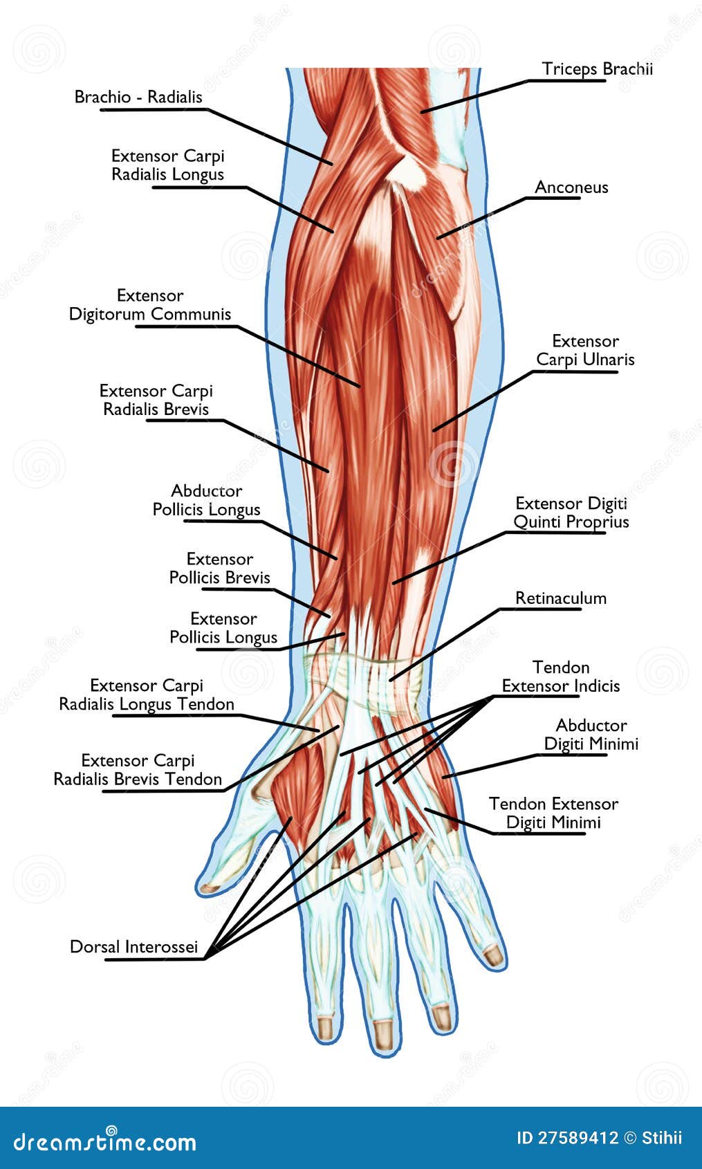
Anatomy Of Muscular System Hand Forearm Palm M Stock Illustration Illustration Of Athletic Health 27589412 from thumbs.dreamstime.com The main knuckle joints are formed by the connections of the phalanges to the metacarpals. The tendon travels along the inside of the forearm on the side of the small finger and crosses the wrist. Over 100 ligaments and tendons; Its muscle belly is in the forearm. See full list on eorthopod.com The mcp joints work like a hinge when you bend and straighten your fingers and thumb. The hand also must be coordinated to perform fine motor tasks with precision. Adequate strength forms the basis for normal hand function.
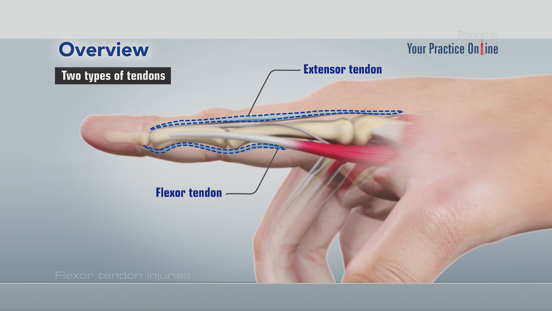
When fingers joints straighten, they are being pulled by the extensor tendons.
What is the function of tendons in the hand? Adequate strength forms the basis for normal hand function. It attaches to the base of the second and third hand bones. The wrist itself contains eight small bones, called carpals. They are controlled by muscles in the forearm. The structures that form and move the hand require proper alignment and control in order for normal hand function to occur. The tendon travels along the inside of the forearm on the side of the small finger and crosses the wrist. Few structures of the human anatomy are as unique as the hand. The hand also must be coordinated to perform fine motor tasks with precision. See full list on eorthopod.com Small bone shafts called phalangesline up to form each finger and thumb. Hand a hand is a prehensile multi fingered appendage located at the end of the forearm or forelimb of primates such as humans chimpanzees monkeys and lemurs human anatomy for the artist the dorsal hand the dorsal the easiest tendons to identify in the dorsal hand are those of the extensor digitorum muscle its name means extensor of the digits which is. When our hands are free of problems, it's easy to take the complex anatomy of the hand for granted.
What is the function of tendons in the hand? Adequate strength forms the basis for normal hand function. This is the other tendon that bends the wrist. One metacarpal connects to each finger and thumb. The structures that form and move the hand require proper alignment and control in order for normal hand function to occur.
Flexor Tendon Injuries Video Medical Video Library from www.ypo.education There are five metacarpals forming the palm of the hand. Adequate strength forms the basis for normal hand function. Further into the palm, the carpals connect to the metacarpals. See full list on eorthopod.com Bones and joints there are 27 bones within the wrist and hand. The wrist itself contains eight small bones, called carpals. When our hands are free of problems, it's easy to take the complex anatomy of the hand for granted. The carpals join with the two forearm bones, the radius and ulna, forming the wrist joint.
The carpals join with the two forearm bones, the radius and ulna, forming the wrist joint.
The main knuckle joints are formed by the connections of the phalanges to the metacarpals. It also attaches to the trapezium, one of your wrist bones. Conditions that change the way these structures work can greatly impact whether the hand functions normally. The structures that form and move the hand require proper alignment and control in order for normal hand function to occur. The hand needs to be mobile in order to position the fingers and thumb. Black and white print showing the musculoskeletal system of a human hand, including the bones, muscles, cartilage, tendons, ligaments, and joints,. Close to the flexor retinaculum, this ligament affects muscles that help extend fingers and other parts of the hand. Anatomy •flexor digitorum profundus tendon : The fcu tendon is one of two tendons that bend the wrist. They are controlled by muscles in the forearm. Dec 18, 2017 · there are 6 tendons that help move your wrist. See full list on eorthopod.com When fingers joints straighten, they are being pulled by the extensor tendons.
Sechrest, md narrates an animated tutorial on. See full list on eorthopod.com Bones and joints there are 27 bones within the wrist and hand. See full list on eorthopod.com The hand needs to be mobile in order to position the fingers and thumb.
Hand Pain The Center For Physical Rehabilitation from pt-cpr.com When our hands are free of problems, it's easy to take the complex anatomy of the hand for granted. See full list on eorthopod.com What parts make up the hand 2. The fcu tendon is one of two tendons that bend the wrist. It attaches to the wrist bone, the pisiform, and as well as the 5th hand bone. Small bone shafts called phalangesline up to form each finger and thumb. The mcp joints work like a hinge when you bend and straighten your fingers and thumb. The tendons that run down our fingers are held in place by a series of.
The hand needs to be mobile in order to position the fingers and thumb.
There are five metacarpals forming the palm of the hand. What are the tendons of the hand? Browse 318 hand anatomy tendons stock photos and images available, or start a new search to explore more stock photos and images. More images for tendon diagram of hand » Sechrest, md narrates an animated tutorial on. The tendons that run down our fingers are held in place by a series of. Its muscle belly is in the forearm. The hand is formed of numerous structures that have an important role in normal hand function. Over 100 ligaments and tendons; What is the function of tendons in the hand? In this image, you will find ulnar artery, ulnar nerve, common palmar digital branches of median nerve, hypothenar muscles, common flexor sheath, ulnar bursa, 5th finger tendinous sheath, synovial tendinous sheath, midpalmar space, insertion of flexor digitorum superficialis tendon, insertion of flexor. It attaches to the base of the second and third hand bones. The fcu tendon is one of two tendons that bend the wrist.


0 Komentar